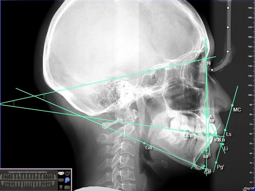
In the early 1950’s, Dr. Steiner emphasized the significance of the cephalometric analysis since the cephalometer was still starting to gain popularity during his time. He tried to present the analysis in an easily understood and relevant manner to encourage higher degree of acceptance of orthodontists to do cephalometric analysis as they manage patients. The tracings were simplified and the methods used gave upfront values for the assessed areas of the head. Throughout the years, Steiner’s analysis has been generally the most commonly used cephalometric analysis. It was published in 1953, providing a series of measurements essential for diagnosis and treatment planning. It focused on the three areas of concern: skeletal, dental and soft tissue. Skeletally, the maxilla and mandible are related to the cranial base (SN) through SNA, SNB, ANB, occlusal plane and mandibular plane (GoGn). The dental values are namely the maxillary incisor position, mandibular incisor position, interincisal angle and lower incisor to chin. These values relate the upper and lower incisors to each other and to the upper and lower dental bases. The soft tissue value, particularly the Steiner’s line (S line), measures the upper and lower lip protrusion. The S line runs from the soft tissue contour of the chin to the tip of the nose.
Steiner identified a difficulty of identifying the porion point since it is a point upon the opening of the auditory canal. The tracing is made from the tip of the ear posts and not on the porion point itself. The Sella-Nasion plane was the baseline for Steiner’s analysis since it is located in the midsagittal plane and is easily located on the xray. The angles SNA and SNB show the amount of protrusion or retrusion of the maxilla and mandible respectively. The angle ANB, which is the difference between SNA and SNB, gives the relationship of the chin to other facial structures. The axial inclination of upper incisor is related to the SN line. The upper first molar is also related to the line NA through locating the most mesial point on its crown to NA. This is important to determine the molar anteroposterior movement. The point A is the deepest point on the contour of the premaxilla. The point B is the deepest point on the contour of the mandible. The most anterior part of the symphysis of the mandible is noted as the pogonion while the most inferior portion is labeled as menton. The gonion is a constructed point located by two tangents, one on the infero-posterior border of the mandible and the posterior border of the ramus.The Gonion-Gnathion (GoGn) line represents the body of the mandible since it closely represents the mass of the body of the mandible and not its inferior border. The lower central incisors’ axial inclination is related to the mandibular plane and so is the upper central incisors to the maxillary base plane. At the end of landmark identification and tracing of line segments relating to each other in angles, is the interpretation of data. From the ANB, the patient’s malocclusion may be classified as Class I, II or III. While the interincisal angle relates the upper and lower incisors to serve as a guide to the orthodontist which torque/prescription is the most appropriate to aim at for orthodontic correction. Despite the challenges encountered with radiograph tracing such as distortion and superimposition of the left and right sides of the skull, it should be remembered that the changes in the position of the landmarks should be expressed in relation to another plane or a reference point.
Steiner’s analysis has proven to be a very useful tool for the orthodontist by being simplified yet relevant not just to diagnosis but also to proper treatment planning.